
Reduced deaf1 mRNA expression during STZ-induced diabetes mellitus inhibits foxp3+regulatory T-cells differentiations in rat’s pancreatic lymph nodes
Abstract
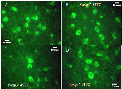
We studied to determine the effect of the levels of Deaf1 mRNA expression on the nature of Foxp3+ Treg cells differentiation during experimental STZ-induced diabetes mellitus (EDM) in rats PLN. To determine the level of Deaf1 mRNA expression was performed RT-PCR in real-time by thermocycler CFX96â„¢ Real-Time PCR Detection Systems. The Foxp3+-immunopositive lymphocytes were determined using an indirect immunofluorescence technique with using a monoclonal rat antibody. We established that development of EDM was accompanied by decreased the expression levels of the transcriptional regulator Deaf1 4,2â€fold in rats PLN with 3-week EDM and 2,5â€fold in rats with 5-week EDM. Reduced Deaf1 mRNA expression during EDM associated with an decreased of total amount of Treg in the PLN, led to changes of distribution into individual classes of FoxP3+ lymphocytes and FoxP3 concentration in immunopositive cells.
Â
Full Text:
PDFReferences
Jeker L, Bour-Jordan H, Bluestone J. Breakdown in peripheral tolerance in type 1 diabetes in mice and humans. Cold Spring Harb Perspect Med. 2012; 2(3):a007807.
Metzger T, Anderson M. Control of central and peripheral tolerance by Aire. Immunol Rev. 2011; 241(1):89-103.
Eldershaw S, Sansom D, Narendran P. Expression and function of the autoimmune regulator (Aire) gene in non-thymic tissue. Clin. Exp. Immunol. 2011; 163:296-308.
Gardner J, Devoss J, Friedman R. Deletional tolerance mediated by extrathymic Aire-expressing cells. Science. 2008; 321:843-847.
Cohen J, Tewalt E, Rouhani S. Tolerogenic properties of lymphatic endothelial cells are controlled by the lymph node microenvironment. PLoS ONE. 2014; 9:e87740
Fletcher A, Malhotra D, Turley S. Lymph node stroma broaden the peripheral tolerance paradigm. Trends Immunol. 2011; 32:12-18.
Fletcher A, Lukacs-Kornek V, Reynoso E. Lymph node fibroblastic reticular cells directly present peripheral tissue antigen under
steady-state and inflammatory conditions. J. Exp. Med. 2010; 207:689-697.
Jensik P, Huggenvik J, Collard M. Identification of a nuclear export signal and protein interaction domains in deformed epidermal autoregulatory factor-1 (DEAF-1). J. Biol. Chem. 2004; 279:32692-32699.
Chow Z, Banerjee A, Hickey M. Controlling the fire - tissue-specific mechanisms of effector regulatory T-cell homing. Immunol. Cell Biol. 2015; 93: 355-363.
Tan T, Xiang Y, Chang C, Zhou Z. Alteration of regulatory T cells in type 1 diabetes mellitus: a comprehensive review. Clin. Rev. Allergy Immunol. 2014; 47(2): 234-243.
Calderon B, Unanue E. Antigen presentation events in autoimmune diabetes. Curr Opin Immunol. 2012; 24: 119-128.
Gagnerault M, Luan J, Lotton C, Lepault F. Pancreatic lymph nodes are required for priming of β cell reactive T cells in NOD mice. J Exp Med. 2002; 196:369-377.
Levisetti M, Suri A, Frederick K, Unanue E. Absence of lymph nodes in NOD mice treated with lymphotoxin-β receptor immunoglobulin protects from diabetes. Diabetes. 2004; 53: 3115-3119.
Malhotra D, Fletcher A, Astarita J. Immunological genome project consortium. Transcriptional profiling of stroma from inflamed and resting lymph nodes defines immunological hallmarks. Nat. Immunol. 2012; 13:499-510.
Yip L, Fuhlbrigge R, Taylor C. Inflammation and hyperglycemia mediate Deaf1 splicing in the pancreatic lymph nodes via distinct pathways during type 1 diabetes. Diabetes. 2015; 64(2):604-617.
Xing Y, Hogquist K. T-cell tolerance: central and peripheral. Cold Spring Harb Perspect Biol. 2012;4
Lee J, Epardaud M, Sun J, Becker J, Cheng A, Yonekura A, Heath J, Turley S. Peripheral antigen display by lymph node stroma promotes T cell tolerance to intestinal self. Nat. Immunol. 2007; 8(2):181-190.
Magnusson F, Liblau R, von Boehmer H, Pittet M, Lee J, Turley S, Khazaie K. Direct presentation of antigen by lymph node stromal cells protects against CD8 T-cell-mediated intestinal autoimmunity. Gastroenterology. 2008; 134:1028-1037.
Yip L, Su L, Sheng D, Chang P, Atkinson M, Czesak M, Albert P, Collier A, Turley S, Fathman C, Creusot R. Deaf1 isoforms control the expression of genes encoding peripheral tissue antigens in the pancreatic lymph nodes during type 1 diabetes. Nat Immunol. 2009; 10:1026-1033.
Yip L, Fathman C. Type 1 diabetes in mice and men: gene expression profiling to investigate disease pathogenesis. Immunol Res. 2014; 58:340-350.
Yip L, Creusot R, Pager C, Sarnow P, Fathman C. Reduced DEAF1 function during type 1 diabetes inhibits translation in lymph node stromal cells by suppressing Eif4g3. J. Mol. Cell Biol. 2013; 5:99-110
Ferraro A, Socci C, Stabilini A, Valle A, Monti P, Piemonti L. Expansion of Th17 cells and functional defects in T regulatory cells are key features of the pancreatic lymph nodes in patients with type 1 diabetes. Diabetes. 2011; 60:2903-2913.
Nti B, Markman J, Bertera S, Styche A, Lakomy R, Subbotin V. Treg cells in pancreatic lymph nodes: the possible role in diabetogenesis and beta cell regeneration in a T1D model. Cell Mol. Immunol. 2012; 9: 455-463.
Tonkin D, Haskins K. Regulatory T cells enter the pancreas during suppression of type 1 diabetes and inhibit effector T cells and macrophages in a TGF-beta-dependent manner. Eur J Immunol. 2009; 39: 1313-1322.
Willcox A, Richardson S, Bone A, Foulis A, Morgan N. Analysis of islet inflammation in human type 1 diabetes. Clin Exp Immunol. 2009;155: 173-181.
Yang S, Fujikado N, Kolodin D, Benoist C, Mathis D. Immune tolerance. Regulatory T cells generated early in life play a distinct role in maintaining self-tolerance. Science. 2015; 348(6234):589-594.
Gardner J, Metzger T, McMahon E. Extrathymic Aire-expressing cells are a distinct bone marrow-derived population that induce functional inactivation of CD4⺠T cells. Immunity. 2013; 39:560-572.
Sun J, Fu H, Wu J, Zhu W, Li Y, Yang W. Macrophages overexpressing Aire induce CD4+Foxp3+ T cells. Mol Med Rep. 2013; 7(1):159-165.
Hammerschmidt S, Ahrendt M, Bode U, Wahl B, Kremmer E, Förster R, Pabst O. Stromal mesenteric lymph node cells are essential for the generation of gut-homing T cells in vivo. J. Exp. Med. 2008; 205:2483-2490.
Rouhani S, Eccles J, Tewalt E, Engelhard V. Regulation of T-cell tolerance by lymphatic endothelial cells. J. Clin. Cell Immunol. 2014; 5. pii: 1000242.
Dubrot J, Duraes F, Potin L, Capotosti F, Brighouse D. Lymph node stromal cells acquire peptide-MHCII complexes from dendritic cells and induce antigen-specific CD4+ T cell to lerance. J. Exp. Med. 2014; 211: 1153-1166.
Refbacks
- There are currently no refbacks.